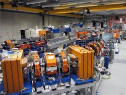
Nijmegen and Amsterdam, The Netherlands--Chemists from Radboud University Nijmegen and the Foundation for Fundamental Research on Matter (FOM) have produced detailed 3D structures of selected peptides (the building blocks of proteins) using far-infrared light produced by Radboud University Nijmegen’s FELIX laser.1 They used vibrational spectroscopy along with harmonic discrete Fourier-transform (DFT) calculations that allowed them to extract the peptides' structural information from the resulting very complex spectra.
Light with wavelengths of up to 100 microns was used. The free-electron laser in the FELIX lab at Radboud University Nijmegen is one of the few lasers in the world capable of directly producing such light. "What is new about our far-infrared spectrum is that we have obtained insights in the folding of the peptides in much more detail, as well as which interactions play a role in this," says Anouk Rijs, assistant professor at the Department of Molecular and Biophysics at Radboud University Nijmegen. "This is important, for instance, for research into interactions between medicines and molecules in the body. Without far-infrared light it is not possible to obtain such detailed information about the molecular structure."
Theories inadequate
Chemists normally determine the molecular structure by comparing the measured infrared spectrum with theoretically predicted spectra. However, the new method produced so much more detail that the old theories and mathematical models were inadequate. "We therefore needed to develop new methods, which we did with colleagues from France," says Rijs. "Using all this new information, we were able to produce an accurate 3D model of the peptides. The next step will be to study larger and more complex molecules."
Source: http://www.ru.nl/english/university/vm/news/@934025/peptides-imaged-in-u/
REFERENCE:
1. Sander Jaeqx et al., Angewandte Chemie (2014) doi: 10.1002/anie.201311189

