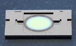
Dresden, Germany--Conventional means to treat brain swelling in cranial injury and stroke victims is to use a cylindrical trephine drill to cut the cranial bone to release pressure in a procedure called a release craniotomy. But because trephines can injure the meninges or outermost layer of the brain (http://www.laserfocusworld.com/articles/2011/01/optogenetics-turning-light-bulbs-on-in-the-brain.html) and possibly lead to meningitis, researchers at the Fraunhofer Institute for Photonic Microsystems IPMS in Dresden together with colleagues at the Fraunhofer Institute for Laser Technology ILT (Aachen, Germany) and at the Fraunhofer Institute for Integrated Circuits IIS (Erlangen, Germany) intend to lower this risk by replacing the trephine with a high-energy femtosecond laser (http://www.laserfocusworld.com/articles/2012/12/fs-laser-surgery-calmar-cavitation-bubbles.html).
"Our colleagues at Fraunhofer ILT have engineered a device that allows the surgeon to guide the laser beam and cut the cranial bone," says Thilo Sander, group manager at Fraunhofer IPMS. The laser beam is fed into the handpiece through an articulated mirror arm. Its core consists of two new types of micromirrors that the researchers at Fraunhofer IPMS developed. The first makes the cranial vault incision; it directs the laser beam dynamically across the cranial bones. The second adjusts any malpositioning.
The process was made possible by miniaturizing the components all while making them able to tolerate up to 20 W of laser output--about 200X more power than conventional micromirrors that reach their limit at 100 mW or so, depending on their specific design. In addition, at 5 x 7 or 6 x 8 mm, they are very large and can guide large-diameter laser beams. By comparison: Conventional micromirrors measure from 1 to 3 mm.
To achieve this breakthrough in micromirror design, the researchers (unlike the silicon panel in conventional micromirrors that is mirrored by an aluminum layer measuring 100 nm thick) applied highly reflective electric layers to the silicon substrate. In the visible spectral range, the mirror reflects not merely 90% of the laser beam like typical components, but 99.9% instead. Much less of the high-energy radiation penetrates into the substrate, allowing the mirror to tolerate large power levels.
The challenge for the researchers was to fabricate this coating on the silicon substrate, just a few micrometers thin. Because the researchers applied several different layers--altogether a few micrometers thick--in order to achieve the desired reflective properties, the mechanical stress prevalent in each of the layers due to temperature expansion differentials had to be resolved. To prevent substrate deformation or arching that diminishes the optical quality of the mirror, Sander says that the effect was counterbalanced by applying the same coating on the reverse side of the substrate.
Demonstration models of the handpiece and the micromirror already exist and the researchers are exhibiting them at Laser World of Photonics from May 13 to 16 in Munich (Hall B2, Booth 421). In subsequent stages of development, the scientists intend to optimize the incision performance.
SOURCE: Fraunhofer IPMS; http://www.ipms.fraunhofer.de/en/press-media/press/2013/2013-05-03.html

