Lausanne, Switzerland--Two École polytechnique fédérale de Lausanne (EPFL) scientists have developed a device that can create three-dimensional (3D) images of living cells and track their reaction to various stimuli without the use of contrast dyes or fluorophores. Yann Cotte and Fatih Toy have combined holographic microscopy and computational image processing to observe living biological tissues at the nanoscale. Their research is being done under the supervision of Christian Depeursinge, head of the Microvision and Microdiagnostics Group in EPFL's School of Engineering.
Using their setup, 3D images of living cells can be obtained in just a few minutes--instantaneous operation is still in the works--at an incredibly precise resolution of less than 100 nm. And because they’re able to do this without using contrast dyes or fluorescent agents, the experimental results don’t run the risk of being distorted by the presence of foreign substances. "We can observe in real time the reaction of a cell that is subjected to any kind of stimulus," explains Cotte. "This opens up all kinds of new opportunities, such as studying the effects of pharmaceutical substances at the scale of the individual cell, for example."
In Nature Photonics, the researchers demonstrate the potential of their method by developing, image by image, the film of a growing neuron and the birth of a synapse, caught over the course of an hour at a rate of one image per minute. The work was carried out in collaboration with the Neuroenergetics and cellular dynamics laboratory in EPFL's Brain Mind Institute, directed by Pierre Magistretti. "Because we used a low-intensity laser, the influence of the light or heat on the cell is minimal," continues Cotte. "Our technique thus allows us to observe a cell while still keeping it alive for a long period of time."
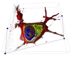
As the laser scans the sample from various angles, numerous images extracted by holography are captured by a digital camera, assembled by a computer and "deconvoluted" in order to eliminate noise. To develop their algorithm, the young scientists designed and built a "calibration" system in the school's clean rooms (CMI) using a thin layer of aluminum that they pierced with 70nm diameter "nanoholes" spaced 70 nm apart. Finally, the assembled 3D image of the cell, that looks as focused as a drawing in an encyclopedia, can be virtually sliced to expose its internal elements, such as the nucleus, genetic material and organelles.
In a company that's in the process of being created and in collaboration with the startup Lyncée SA, they hope to develop a system that could deliver these kinds of observations in vivo, without the need for removing tissue, using portable devices. In parallel, they will continue to design laboratory material based on these principles.
SOURCE: EPFL; http://actu.epfl.ch/news/peering-into-living-cells/
IMAGE:Holographic microscopy combined with computational imaging can see cells in three dimensions without contrast agents. The image can be virtually sliced to expose its internal elements, such as the nucleus, genetic material, and organelles. (Courtesy EPFL)
