RAMAN SPECTROSCOPY/DRUG TESTING: SERS will add real-time analysis to painless microneedle patch for drug testing

Jan. 21, 2014
A bandage-like patch of polymer microneedles, paired with surface-enhanced Raman scattering (SERS), may enable a future method for safe and painless testing for drugs and some infections, along with real-time analysis.

A bandage-like patch of polymer microneedles, paired with surface-enhanced Raman scattering (SERS), may enable a future method for safe and painless testing for drugs and some infections, along with real-time analysis.

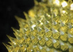
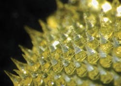
Swollen needles on a used microneedle patch. In testing, patients likened the sensation of the patch being pressed into the skin as an encounter with Velcro.

Experiments conducted at the Queen's University Belfast laboratories of Ryan Donnelly, a researcher in the School of Pharmacy, are showing that the array of tiny polymer needles on the underside of the patch can absorb fluids in the skin's surface such as salts and fatty acids.

According to Donnelly, "we typically find the same compounds in this interstitial fluid as you would find in the blood. But compared with drawing blood, our patches can get their samples in a minimally invasive way. And it's far safer than using a conventional needle. These microneedles, once they have been used, become softened, so that there's no danger of dirty needles transferring infection to another patient or one of the healthcare workers." Two million healthcare workers are infected by needlestick injuries every year.

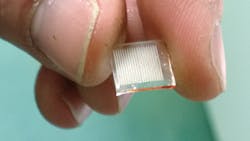
Microneedle patch that could be used for painless drug sampling. (Both images courtesy of Ryan Donnelly, School of Pharmacy, Queen's University Belfast)

The microneedles are made of polymer gel. The backing material can be made chemically attractive to target compounds, encouraging them to diffuse into the gel with interstitial fluid drawn out of the skin and locking them in place for later analysis. Real-time monitoring might involve using SERS to detect drug compounds inside the gel. The group already has proof-of-concept for this idea and is now looking to extend the range of drug concentrations that can be detected in this manner.

The group is currently in discussions with a major medical manufacturer to produce prototype commercial devices, following the first stage of full clinical trials.
Sponsored Recommendations

Melles Griot® XPLAN™ CCG Lens Series

March 19, 2024
IDEX Health & Science sets a new standard with our Melles Griot® XPLAN™ CCG Lens Series fluorescence microscope imaging systems. Access superior-quality optics with off-the-shelf...

Spatial Biology

March 19, 2024
Spatial Biology refers to the field that integrates spatial information into biological research, allowing for the study of biological systems in their native spatial context....

Fluorescent Protein Optical Imaging Considerations

March 19, 2024
What factors should you consider when your incorporate fluorescent proteins in an optical imaging application? Learn more.

Custom-Engineered Optical Solutions for Your Application

March 19, 2024
We combine advanced optical design and manufacturing technology, with decades of experience in critical applications, to take you from first designs to ongoing marketplace success...

Voice your opinion!

To join the conversation, and become an exclusive member of Laser Focus World, create an account today!