Exhibit celebrates 'The Art of Cell Biology'

March 18, 2014
Honoring the work of his cell biology students over the past 10 years, Bowdoin College (Brunswick, ME) professor Bruce Kohorn created an art exhibit showcasing his students' optical microscopy images.
(Images courtesy of Bruce Kohorn/Bowdoin College)
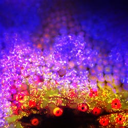
The surface of a leaf captured using single-focal-plane confocal microscopy. Green represents cell-surface green fluorescent protein (GFP); blue, chlorophyll; and red, red lipid membrane dye.

Honoring the work of his cell biology students over the past 10 years, Bowdoin College (Brunswick, ME) professor Bruce Kohorn created an art exhibit showcasing his students' optical microscopy images.

Called "The Art of Cell Biology" and held Jan. 27 through Feb. 7, 2014, at the college's Visual Arts Center, the exhibit's artwork was all developed using compound fluorescence and confocal microscopy, including single-focal-plane and 3D confocal techniques. A National Science Foundation-funded Zeiss LSM 510 Meta system with 488 nm (UV) excitation by an argon laser and 515 nm (green) emission enabled creation of many of the images, Kohorn told BioOptics World.

The students who participated in the exhibit gained an understanding of cell structure and function, Kohorn explained. But the students also saw the images' aesthetic value: Noah Gavil '14 said, "You definitely have your moments when you just stop thinking about what you're actually studying and looking for and just think, 'That's awesome; that's a gorgeous picture.'"

To view the full exhibit, please visit www.bowdoin.edu/faculty/b/bkohorn/images/gallery/index.shtml.
About the Author

Lee Dubay | Managing Editor

Lee Dubay is managing editor for Laser Focus World. She is a seasoned editor and content manager with 20 years of experience in B2B media. She specializes in digital/print content management, as well as website analytics, SEO, and social media engagement best practices. 

Sponsored Recommendations

Melles Griot® XPLAN™ CCG Lens Series

March 19, 2024
IDEX Health & Science sets a new standard with our Melles Griot® XPLAN™ CCG Lens Series fluorescence microscope imaging systems. Access superior-quality optics with off-the-shelf...

Spatial Biology

March 19, 2024
Spatial Biology refers to the field that integrates spatial information into biological research, allowing for the study of biological systems in their native spatial context....

Fluorescent Protein Optical Imaging Considerations

March 19, 2024
What factors should you consider when your incorporate fluorescent proteins in an optical imaging application? Learn more.

Custom-Engineered Optical Solutions for Your Application

March 19, 2024
We combine advanced optical design and manufacturing technology, with decades of experience in critical applications, to take you from first designs to ongoing marketplace success...

Voice your opinion!

To join the conversation, and become an exclusive member of Laser Focus World, create an account today!