Infrared (IR) light—which is invisible to the human eye—has broad application in many areas because its energy, in the form of heat, is easiest to discern. When something is not hot enough to emit visible light, it emits most of its energy in the IR.
It is important to note that IR light, like the visible light region, contains ranges of wavelengths, each with different properties. Here, we will focus on the near-infrared (NIR), the range closest to visible red light, which is effective for the life sciences. I spoke with three NIR light source makers to learn more about their advantages in bio, the different types of available sources, and what's in store for the future.
NIR: The sweet spot for bio
NIR light, which ranges from 750 to 2500 nm, is used in several life science and biomedical applications because it exhibits less intrinsic absorption and inherently less scattering than using ultraviolet (UV) and visible light. "Because biological samples are complex, optical measurements using UV and visible illumination are hindered by absorbance, fluorescence, and scattering of light by various sample constituents," explains Yvette Mattley, Ph.D., senior applications scientist at Ocean Optics (Dunedin, FL). So NIR illumination facilitates the optical characterization of specimens with less complicated, more accurate data, she adds.
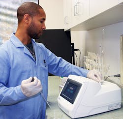
Life science and biomedical applications that benefit from NIR light are plentiful, particularly in measurement applications such as microscopy, optical coherence tomography (OCT), and spectroscopy (see Fig. 1). "These measurements provide a wealth of information on sample characteristics, including chemical composition, localized analysis of fluorophore tags, biomolecule aggregation and diffusion, cell structure, and tissue composition," says Mattley. In microscopy, longer NIR wavelengths enable deeper sampling depths in-vivo and in-vitro due to low absorption and scattering. Using NIR excitation sources in vibrational spectroscopy on organic samples is beneficial because organic materials tend to have a high degree of autofluorescence when excited with visible or UV radiation, says Robert Chimenti, marketing manager at B&W Tek (Newark, DE). He adds that another common application that utilizes NIR light is laser medicine, particularly aesthetics, where wavelengths between 750 and 1100 nm are preferable due to the high transmissivity of oxyhemoglobin and water when compared to melanin and other pigments, which are highly absorbent in the same spectral region.
The benefits don't end there, either. Mattley adds that NIR light is typically nondestructive when characterizing tissue and biological samples, which is key for noninvasive clinical work.
Light source types
Right now, there are NIR light sources available for almost any life science application, ranging in capability and cost: NIR lasers, NIR laser diodes, NIR LEDs, incandescent sources filtered for NIR, and supercontinuum lasers.
"NIR LEDs are often used as a low-cost alternative for applications that do not require broad spectrum or high brightness," explains Chimenti, including low-level light therapy. High-brightness LEDs, such as Ocean Optics' LLS series, provide narrow bands of NIR light for excitation of fluorescent probes used to label cellular constituents or other biomolecules.
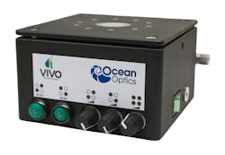
For applications requiring broad spectrum illumination, broadband tungsten-halogen sources provide illumination from the visible region through the NIR for absorbance, transmission, and reflectance measurements, says Mattley. The company's Vivo direct illumination source performs NIR diffuse reflection measurements of optically dense biological, agricultural, and pharmaceutical samples, she adds (see Fig. 2). NIR tunable lasers enable pulsed or continuous-wave light from visible to NIR wavelengths for fluorescence applications requiring greater imaging depths, such as multiphoton imaging, which benefit from NIR wavelengths for deep in-vivo imaging. The InSight DeepSee ultrafast laser from Newport (Irvine, CA), for instance, has a 680–1300 nm tuning range and ultrashort, 100 fs pulse widths for exciting multiple fluorophores of interest. And supercontinuum lasers, such as the SuperK supercontinuum white light laser from NKT Photonics (Birkenrød, Denmark), "emit light in the entire 400–2400 nm wavelength range, thereby covering most of the important wavelengths for life science," explains Kim P. Hansen, global product line manager, marketing at NKT Photonics. For example, these sources are being used with Gaussian-shaping filters to provide a Gaussian spectral output with a width of several hundred nanometers ideal for OCT, where the broad spectral illumination results in resolution matching that of titanium sapphire (Ti:sapphire) systems at much lower cost, she adds.Catering to life science applications requiring high brightness and broad spectrum illumination, Chimenti mentions laser-driven light sources (LDLS), a recent development by Energetiq (Woburn, MA), in which a laser-driven, light-emitting plasma produces extremely high brightness and a flat spectral profile over the entire region, enabling coupling into small core fibers and imaging onto small samples.
A bright future
"Continuing advances in LED technology will be used to create more powerful, narrower band, and novel excitation sources for fluorescence," says Mattley. Also, she and Chimenti agree that the future for NIR illumination is higher performance in a smaller, more flexible footprint that's portable and more user-friendly.
Multimodal approaches—which combine two processes to obtain accurate results—have made their mark in life science applications, and will likely benefit from NIR sources. To that end, Chimenti mentions that combining laser surgery with Raman spectroscopy could determine properties of the tissue being operated on, which would mostly likely be performed in the NIR region. And using supercontinuum sources in ophthalmology (retinal imaging) and OCT, for example, could enable users to use both modalities to obtain the maximum amount of information because they emit light across the entire wavelength spectrum.


