MULTIPHOTON MICROSCOPY: Miniature microscope images active neurons
Observing neural activity in the mammalian brain with multiphoton microscopy allows researchers not only to locate individual active neurons, but also to see how groups of neurons coordinate their activity. There’s a problem though: multiphoton microscopes are large. They’re far too large to mount on a test rat, so instead, the rat must be mounted to the microscope—which means that the rat is not moving around in a neuron-activating manner. “Virtual reality” (in other words, placing a video screen in front of the test rat) can help, but only to a limited extent.
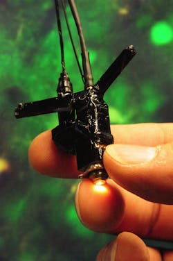
So scientists at the Max Planck Institute for Biological Cybernetics (Tübingen, Germany) and the Max Planck Institute for Medical Research (Heidelberg, Germany) have created a miniature optical-fiber-fed two-photon microscope lightweight enough (5.5 g) to be fastened onto a rat, with the mounting rigid enough that the neurons in the field of view move less than 3 µm relative to the microscope (see figure).1 The microscope is stable enough that images of neurons in a rat’s brain can be recorded for five minutes or more at a time, even as the rat is quickly running about.
Microscopic details
A titanium:sapphire ultrafast laser provides excitation pulses at either 880 or 925 mm, with average powers of 2.4 and 1.6 W respectively. The pulses are brought to the microscope via a single-mode excitation fiber; the researchers tested three types of single-mode fibers to see which one was optically the most stable when flexed, finally settling on a polarization-maintaining fiber (one of the other fibers–a bend-insensitive fiber–showed changes in output polarization as it was flexed).
The microscope has a custom-designed water-immersion lens with a 0.9 numerical aperture, a 0.7 mm focal length, and a working distance of 0.7 mm. The fiber is scanned via a piezoelectric lever in a nonresonant design that provides random access and much more flexibility in its scan patterns than a resonant-scanning design. A plastic optical fiber with a 0.98 mm diameter serves as the collection fiber.
The researchers first tested the microscope on anesthetized animals. The head-mounted microscope was positioned directly over the neuronal area responsive to visual stimuli directly in front of a rat’s nose. The average power of the excitation-laser light exiting the objective was 100 to 150 mW; at the focus it was about 40 to 60 mW, assuming a 200 µm scattering length. No cell damage was observed over a one-hour time period. Moving-grating images placed in front of the rat evoked neuronal responses in the rat as expected.
Next, the microscope was tested with rats that were fully awake and unrestrained. Even in experiments lasting for more than four hours, the rats appeared to be moving around normally, doing the things that rats do: running, digging, feeding, and pouncing; their behavior, as well as their running speeds and accelerations, were similar to rats without microscopes mounted on their heads.
Seeing the brain’s complexity
The neuronal images tracked the activity of astrocytes as well as neurons; astrocytes are believed to be active participants in the brain’s activity, and are a focus of current research.
Additional experiments were carried out, including monitoring of rats as they moved along a semicircular track, and an examination of the relationship between a rat’s shifting gaze and the simultaneously imaged stimulus-evoked neuronal activity.
Occasionally (around 2% of the time), such as when a rat shook its head vigorously, the image was unstable. Nevertheless, the researchers foresee their miniature microscope being especially useful in monitoring brain behavior during such complex tasks as social interactions, capture of prey, and usage of the vestibular system (sense of balance).
REFERENCE
- Jürgen Sawinski et al., PNAS Early Edition, www.pnas.org_cgi_doi_10.1073_pnas.0903680106 (2009).
About the Author

John Wallace
Senior Technical Editor (1998-2022)
John Wallace was with Laser Focus World for nearly 25 years, retiring in late June 2022. He obtained a bachelor's degree in mechanical engineering and physics at Rutgers University and a master's in optical engineering at the University of Rochester. Before becoming an editor, John worked as an engineer at RCA, Exxon, Eastman Kodak, and GCA Corporation.