MICROSCOPY: Confocal microscopy sees large surface area
Minimally invasive microscopy techniques can reduce the need for excisional biopsy, particularly if the technique can image a large tissue area. While reflectance confocal microscopy has been demonstrated in patients, the field of view remains limited. But researchers at Massachusetts General Hospital (Boston, MA) have demonstrated a new spectrally encoded confocal-microscopy (SECM) method that images areas three to four orders of magnitude larger than that of single images obtained by conventional confocal microscopes.1
The SECM device was designed to image the interior walls of a cylinder with a length of 2.5 cm and a diameter of 2.0 cm—the rough dimensions of the distal human esophagus. In the experimental setup, light from a fiber-coupled 2.0 mW superluminescent diode, centered at 793 nm with a bandwidth of 44 nm, is sent through a 50/50 single-mode fiber-optic splitter. The light is collimated using free-space optics and transmitted axially through the cylinder to a diffraction grating/lens pair that directs the light onto the sample, while the reflected light from the sample is transmitted back through the probe optics.
Helical scanning
The 1780 line/mm diffraction grating, from Holographix (Hudson, MA), and the aspheric imaging lens produces a 500 µm longitudinal array of focused, spectrally encoded spots on the interior wall of the cylindrical sample. This grating/lens pair is attached to the shaft of a 15-mm-diameter motor by a custom-machined housing. As the motor rotates, a spectrally encoded line is scanned across the inner circumference of the sample. To increase the imaging area, the motor is also translated across the sample, allowing the acquisition of a helical scan of the entire cylindrical interior of the sample.
A custom-built spectrometer and linear CCD collects the helical scan data. The system digitizes approximately 60,000 points per motor rotation (30 rpm) to achieve 1 µm circumferential sampling. The time required for one complete scan through the 2.5-cm-long cylindrical sample was 100 seconds, with a motor translation speed of 0.25 mm/s. With an effective numerical aperture of 0.4 for the scanning system, theoretical transverse and axial resolutions were computed to be 1.0 and 3.6 µm, respectively. Assuming aberration-free operation, the theoretical spectral resolution on the sample was 0.08 nm with a maximum of approximately 630 resolvable points across the spectrally encoded 500 µm line.
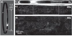
The researchers tested their confocal microscopy system on a U.S. Air Force resolution chart mounted on a piece of lens paper affixed to the interior of a cylinder, as well as on a swine intestinal-tissue fragment. For the swine intestinal tissue, images were displayed at magnifications of 1×, 4×, 20×, and 40× (see figure). Magnified images of the tissue demonstrate features consistent with intestinal villi, containing bright reflective dots that may be nuclei or intracellular vacuoles.
According to researcher Dvir Yelin, several challenges remain before the SECM technique can be applied in a clinical setting. First and foremost, miniaturization of the optics, motor, and housing is required. A method for precise centering of the imaging optics needs to be developed, along with faster data acquisition and processing. “Once these technical issues are resolved, the acquired microscopic data sets will be huge,” says Yelin. “In order to manage this information, we will additionally have to develop a user interface, where a physician can examine the dataset at low power, and gradually zoom in to specific sites to inspect cellular and subcellular structures.”
Guillermo Tearney, a pathologist and senior author on the paper, believes that this technique may usher in a new paradigm for medical diagnosis. “Instead of excisional biopsies, which often represent only small portions of the area of interest, this technique could significantly increase diagnostic yield by rapidly and noninvasively imaging an entire epithelial surface with microscopic resolution,” says Tearney.
REFERENCE
1. D. Yelin et al., Optics Lett. 32(9) 1102 (May 1, 2007).
About the Author

Gail Overton
Senior Editor (2004-2020)
Gail has more than 30 years of engineering, marketing, product management, and editorial experience in the photonics and optical communications industry. Before joining the staff at Laser Focus World in 2004, she held many product management and product marketing roles in the fiber-optics industry, most notably at Hughes (El Segundo, CA), GTE Labs (Waltham, MA), Corning (Corning, NY), Photon Kinetics (Beaverton, OR), and Newport Corporation (Irvine, CA). During her marketing career, Gail published articles in WDM Solutions and Sensors magazine and traveled internationally to conduct product and sales training. Gail received her BS degree in physics, with an emphasis in optics, from San Diego State University in San Diego, CA in May 1986.