OPTICAL-COHERENCE TOMOGRAPHY: Time-domain OCT brings new vision to dental diagnostics
Studies have shown that near-infrared (NIR) imaging in the 1300 nm range offers several advantages over conventional diagnostic tools used in dentistry, such as x-rays, particularly for early detection of occlusal and interproximal dental decay and for identifying decay hidden under tooth enamel, which often goes unseen on x-rays (see www.laserfocusworld.com/articles/292402 and www.laserfocusworld.com/articles/192733). So far, however, there has been no commercially available dental-imaging system that utilizes lasers or superluminescent diodes in the NIR.
One company is hoping to change this by introducing the first optical-coherence-tomography (OCT) system for dental imaging, in 2008. Utilizing proprietary technology plus licensed OCT methodologies from Lawrence Livermore National Laboratory (Livermore, CA) and Lightlab Imaging (Westford, MA), Lantis Laser (Denville, NJ) has been developing and testing its OCT Dental Imaging System on ex vivo teeth for four years. Now the company is preparing to place five prototypes in universities and private dental practices for further clinical testing, this time in the mouth.
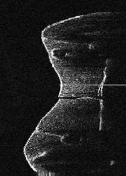
The Lantis OCT system comprises a 1310 nm superluminescent-diode (SLD) light source (20 mW), standard Michelson interferometer, fiber-optic image probe (120 µm diameter), analog image sensor, and digital computer system for image processing and display. It is designed to be used chair-side in the dental examination room, providing real-time images of teeth for the diagnosis of early and secondary caries and subenamel decay (see figure). In operation, the dentist holds the probe up to 3 mm away from the tooth to be imaged. Output from the SLD is split at the fiber-optic coupler and directed toward the sample and reference arms. Reflections from the mirror and backscattered light from the sample are recombined at the coupler and propagated to the detector and light source. A cross-sectional image is produced by transversely scanning the beam across the sample and collecting a reflectance profile at each point.
“We image at a total depth of 7 mm, 3 to 4 mm away from the tooth and 3 mm into the tissue,” said Stan Baron, president and CEO of Lantis. “There are challenges with hard vs. soft tissue; when we image into the occlusal (the molars), there are peaks and valleys, but by holding the scanner tip away from the actual tissue you can still get the depth penetration. And we can do this up to a width of about 10 mm.”
Each image is actually a combination of thousands of points of light gathered in a single scan. In a standard 10 mm scan, the scanning system will stop 4000 times along the 10 mm scan line, taking 2000 measurements at each point at up to 3 mm into the tooth. The result is eight million pieces of information per scan. And all of this happens in a matter of microseconds, according to Baron.
Time-domain vs. Fourier-transform
While Fourier-transform OCT offers advantages over time-domain OCT—including the ability to do three-dimensional imaging—Baron said the company made a conscious decision to go with time-domain OCT to keep the overall cost of the system down and thus make it accessible to most dentists, while still providing sufficient resolution to characterize dental tissue and diseases. The dental market is very price sensitive when it comes to the technologies it will invest in, and early on Lantis set a price cap of $20,000 to $25,000 for the OCT imaging system. While the price is higher than conventional or digital x-ray systems or the laser-based Diagnodent—which uses a 630 nm diode laser to provide fluorescence measurements in fissure areas of the tooth to distinguish between decayed and healthy dentin—Baron believes that the advantages offered by OCT will make the Lantis product “hard to resist.”
“One thing clinicians are looking for in an imaging system is both quantitative and qualitative information, particularly at an early stage. They want to know how much of something there is and where it is, and this is what OCT provides,” Baron said.
While the Diagnodent provides an indication on a scale of 1 to 10 of how much decay is present (via the fluorescence), there has to be a minimum amount of decay before it can detect it, he adds.
“Our OCT system will pick up the presence of decay at a much earlier stage, and it provides a histologic image that indicates both how much decay there is and where it is located, not just a general auditory signal that indicates the presence of some decay.”
At the same time, the fact that dentists have embraced the Diagnodent is a good sign because it means they want, and are ready to invest in, better diagnostic aids, according to Baron. Toward that end, the company is looking at additional applications, including imaging periodontal ligament attachments (measuring the depth of the pockets around teeth) and detecting oral cancers.
“Dentists have been operating ‘blind’ for a long time, which makes diagnosing many clinical conditions very difficult,” said Baron. “We think once they see what OCT can do to help them evaluate dental tissue, it will be very difficult to resist the deployment of OCT in their practice.”
About the Author
Kathy Kincade
Contributing Editor
Kathy Kincade is the founding editor of BioOptics World and a veteran reporter on optical technologies for biomedicine. She also served as the editor-in-chief of DrBicuspid.com, a web portal for dental professionals.