OPTICAL MATERIALS: Compaction and rarefaction affect photolithography system lifetimes
J. MARTIN ALGOTS, CARL STEINBRECHER, SOUPAYA CHUCKRAVANEN, HIROKI JINBO
During the past 10 years, deep-ultraviolet excimer-laser-based systems emerged as the preferred light sources for manufacturing semiconductors. Krypton fluoride (KrF) sources operating at 248 nm have become the production workhorse, and the continuing need for smaller feature sizes has driven development of argon fluoride (ArF) excimer sources operating down to 193 nm as the next production generation. Early development of 193-nm lithography was fueled by 10- and 20‑W sources, but output powers of 40, 60, and eventually 90 W will likely be required to support techniques such as immersion. Such high power levels, of course, call for careful evaluation and testing of core optical materials that are at the heart of modern photolithography systems.
Stringent performance requirements severely limit the number of optical materials that can be used at 193 nm to make the very large, multielement scanner lenses currently used in semiconductor-manufacturing production equipment. The majority of the 20 to 30 lens elements that comprise a modern scanner lens system are based on fused silica. Calcium fluoride (CaF2) is used for some of the smaller elements because of its natural resistance to optical damage under intense ultraviolet radiation but high cost, short supply, complex optical properties, as well as lens manufacturing difficulty prevent widespread adoption of CaF2 in scanner projection optics.
The high energy of 193-nm photons can cause the optical properties of fused silica to change with time, beyond the tight specifications needed to successfully image features below 90 nm. This change can limit the useful life of scanner lenses and illuminator optics. Given the high cost of these lenses, it is beneficial to understand and model the causes of this change. The two most significant mechanisms that affect the index of refraction are compaction and rarefaction (sometimes referred to as decompaction). Cymer has worked with several optical-material and scanner-equipment suppliers to develop models that describe these mechanisms as functions of fluence, number of pulses, and pulse lengths.
null
Test station
To investigate the effects of 193-nm radiation on fused silica, a test station was built to deliver 193-nm radiation at 4 kHz, similar to the illumination in actual scanners, at two pulse lengths and four fluence levels for up to 18 optical test samples. More than 80 billion pulses have been logged on this test station (see Fig. 1).1, 2, 3, 4, 5
The light source runs without bandwidth-narrowing optics (the bandwidth is approximately 300 pm, FWHM), and with output couplers at both ends of the gain region, allowing radiation to be emitted from both ends of the light source. The beam from one end is routed through an optical pulse stretcher to increase the length of the pulse before reaching the test chamber, while the beam from the other end is routed to the test chamber unstretched. Nominal pulse length is approximately 20 ns, while separate tests have been run with the stretched beam at both 40 and 90 ns.
Each of the two beams-stretched and unstretched-from the light source are split into three different sample lines. Each sample line is further split into four more beams, each of which is separately attenuated to produce four different fluence levels per line. The result is that each optical sample will be exposed to four fluence levels, both with the stretched and unstretched beams, resulting in a total of eight exposure regions. Each sample line can accommodate up to six samples illuminated in series.
Each sample is mounted on a linear stage, and a camera and power meter are mounted overhead on an x-y stage to measure the beam before and after each optical sample. A total of 20 linear stages and more than 50 other remote actuators are controlled by two personal computers, which also handle image and data acquisition for the entire test station.
The energy distribution at each sample is determined by imaging the incident and transmitted beam at each optical sample through a custom lens and a cooled CCD camera assembly. Each image is calibrated with a corresponding power-meter reading automatically every 300 million pulses. Pulse lengths are measured with photomultiplier tubes (PMTs) and a high-speed oscilloscope. Pulse lengths and shapes, oxygen content of the test enclosure, shot counts, and other parameters are logged every 15 minutes.
At every 2.5 billion pulses the samples are measured with a Zygo interferometer at 633 nm. The first, unexposed interferometer images are subtracted from the current images and a spot filter is applied to the resultant image. The spot filter subtracts the average of an annular region from the average of a center spot. The spot is slightly smaller than the circular exposure regions and the inner annulus diameter is slightly larger. The annulus outer diameter is chosen so that when the filter is centered on the exposure region, it does not extend off the edge of the sample or onto other exposure regions. The largest deviation of the filtered image for each exposure region is used to calculate the change in index of refraction(δn) at that pulse count.
If the index change in fused silica can be understood and accurately modeled, it may be possible to adjust the light-source beam parameters and the scanner lens materials to produce longer-life scanner lenses. The current model used by Cymer to describe the change of refractive index in fused silica as a function of 193-nm exposure is:
null
where δn is the change in index of refraction, N is the number of pulses, I is the fluence, tis is the length of the individual pulses, and b, k2 Do and ∆nsat are coefficients dependent on the specific fused-silica formulation. The first term of the equation describes the rarefaction and the second term describes the compaction.
Compaction and rarefaction
Compaction in fused silica is the rearranging of the silica molecules in the structure, filling voids in the bulk material formed during manufacture of the fused silica. The number and nature of these voids are determined by the formulation and manufacturing process of the specific fused silica. Compaction results in a more dense material at a lower energy state that affects the optical properties of the material. The energy received from the radiation enables the molecules to move.
The model indicates that this is a two-photon process, that requires the energy of two photons imparted at the same time to allow for the movement of the molecule. The rate at which compaction occurs is a function of the instantaneous energy (fluence2/time [NI2/tis]) of the radiation and the properties of the specific formulation of the fused silica (coefficient k2 and exponent b). Increasing the pulse length and maintaining the same pulse energy should reduce the rate of compaction in a sample.
Rarefaction is the decrease in optical density due to what is believed to be the expansion of the material caused by the formation of SiOH. The hydrogen is introduced in the manufacturing process and can, to some extent, be controlled by the manufacturer. The model indicates that this is a one-photon effect and therefore a function only of the cumulative fluence (NI) and the specific formulation of the fused-silica sample (coefficients ∆nsat and D0). The pulse length should have no effect on rarefaction. If indeed the rarefaction is a function of hydrogen content, the reaction should approach a saturation level (∆nsat) at some continuously decreasing rate (described by the exponential term).
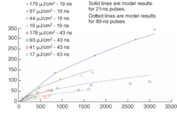
One might imagine that either the compaction or the rarefaction could dominate, depending on the material-dependent constants-that is, in fact, the case. At higher fluence levels, compaction normally dominates and rarefaction was only discovered when exposure at lower fluence levels was investigated. Some fused silica materials will fall quickly into the rarefaction dominated regime at the fluence levels expected in a 193-nm scanner lens, while others move into the compaction-dominated regime (see Fig. 2).
Performance modeling
Once the coefficients for a number of fused-silica materials are determined, the results can be used to model the lifetime effects of radiation on the scanner lenses and illumination optics. The fluences on the lens elements vary with the type of logic layer being exposed, the reticule illumination scheme, and the particular lens element in question. Larger lens elements generally have lower fluences, while smaller elements have larger fluences. Once the fluence levels of each element are determined, the adjustments to the index of refraction can be fed into the lens design programs to determine the effect on the imaging performance of the lens.
Each fused-silica formulation could have a fluence and pulse length that comes close to balancing the rarefaction with compaction. Once the lens designer has determined the fluence levels of each element, different fused-silica formulations can be prescribed for different lens elements to minimize the index changes over the life of the lens. Pulse length is determined by the laser and must be chosen as a global parameter in this very elaborate optimization problem.❏
REFERENCES
1. J.M. Algots et al., Proc. SPIE5040, 1639 (2003).
2. R. Sandstrom, Proc. 2nd Symp. on 193-nm Litho., Colorado Springs, CO (1997).
3. J. Moll, P. Dewa, Proc SPIE4691, 1734 (2002).
4. J.M. Algots et al., Proc. SPIE5377 (2004).
5. R. Morton et al., Proc SPIE4000, 496 (2000).
J. MARTIN ALGOTS is a senior optics engineer, CARL STEINBRECHER is senior manager for direct customer marketing, and SOUPAYA CHUCKRAVANEN is the senior director of direct customer and product marketing at Cymer, 17075 Thornmint Ct., San Diego, CA, 92127; e-mail: [email protected]; www.cymer.com. HIROKI JINBO is the manager of the first development section in the planning and development department of the glass division at Nikon.


