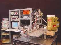
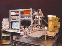
Terahertz technology originally developed at Dartmouth College (Hanover, NH) nearly two decades ago may have finally found its niche—studying proteins and other large molecules with resonances in the terahertz frequency range (see "Old electron microscope generates infrared light," Laser Focus World, October 1998). Licensed to Vermont Photonics (Brattleboro, VT) in 1988, the tunable terahertz-source technology originally devised by Dartmouth physics professor John Walsh has undergone several refinements and improvements, yielding a compact spectroscopic tool, which its developers believe offers some unique capabilities, particularly for molecular biology and proteomics research.
"What we are doing is like a tabletop free-electron laser (FEL) or a nanoklystron that generates a free-space photon field directly," said Mike Mross, president of Vermont Photonics. "With narrow bandwidth, tunability over 0.3 to 3.0 THz, and a 'desktop' footprint, our source offers possibilities for imaging and spectroscopy that are not available using synchrotrons, wiggler FELs, or pulsed terahertz systems."
The original technology licensed from Dartmouth involved a modified scanning-electron microscope (SEM) that yielded far-infrared energy by passing an electron beam over a metal grating, with the electrons being produced by a set of SEM optics. Even then, the wavelength of the source could be tuned from 200 to more than 1000 µm (1.5 to 0.3 THz) with output powers of about a nanowatt. Today the system retains few of its SEM roots, but the underlying configuration is the same: a compact, continuous-wave, narrowband Smith-Purcell tunable terahertz source that now produces several microwatts of power at a bandwidth of around 20 µm with a signal-to-noise ratio of better than 1000:1. Tunability is achieved by adjusting the geometry of the resonance structure or by changing the voltage of the electron beam.
"Continuous-wave powers are in excess of 1 mW and are emitted over a small solid angle from a source area of about 1 mm2," said Thomas Lowell, vice president of Vermont Photonics. "This means that at 400 µm, for example, the source brightness is more than 100 times greater than a mercury arc lamp and about 10 times more than a typical synchrotron source."
Mross and Lowell see multiple molecular research applications for this technology in spectroscopy, imaging, and detection of DNA hybridization states and protein ligand binding states. Research has shown that nanoscale clusters of water molecules support terahertz vibrations and certain organic molecules show strong absorption and dispersion due to rotational and vibrational transitions that are specific to the molecule, thus enabling terahertz fingerprinting. Other potential applications include protein identification for drug discovery, with the terahertz system offering a more-efficient approach to tagging biomarkers.
"It appears that proteins have unique signatures around 1 THz, and researchers are starting to realize that molecules like protein and DNA can resonantly vibrate at frequencies in this range and that those vibrations may actually have something to do with molecular structure and function," Mross said. "While broadband terahertz sources are very good for identifying some protein resonances, you then might want to go in and pump or 'jiggle' these resonances to see how they respond, which is something broadband terahertz cannot do."
Vermont Photonics currently operates two experimental continuous-wave Smith-Purcell tunable terahertz sources at its laboratory. The company is hoping to collaborate with other researchers to begin designing some simple experiments that will demonstrate the utility of this technology for nanoscale research and subcellular function.

