Color-coded quantum dots provide fast DNA testing

Sept. 1, 2001
Indiana University (Bloomington, IN) researchers have developed a multicolor optical-coding method that identifies tens of thousands of genes all at once by using tiny semiconductor crystals that dazzle in ultraviolet light.
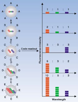
FIGURE 1. In the optical coding method, based on wavelength and intensity multiplexing, the largest spheres represent polymer microbeads, in which smaller colored spheres (quantum dots) are embedded according to predetermined intensity ratios. Molecular probes are attached to the bead surface for biological binding and recognition, such as DNA hybridization. The colored (blue, green, red) spheres illustrate fluorescence intensity levels only. An optical measurement is possible by measuring the fluorescence spectra of single beads.

Indiana University (Bloomington, IN) researchers have developed a multicolor optical-coding method that identifies tens of thousands of genes all at once by using tiny semiconductor crystals that dazzle in ultraviolet light (see Fig. 1). The technique works like a bar codewith each color and intensity combination corresponding to an individual geneand can study up to 40,000 genes or proteins in as little as 10 minutes.

Researchers have tried for many years to use tiny crystals, called quantum dots, as glowing labels for genes, proteins, and other biological molecules. Quantum dots promise to be faster in binding kinetics, more flexible in target selection, and less expensive as tests for on-the-spot biological analysis or patient diagnosis. Until now, they have been difficult to collect and manipulate with enough precision to be useful.

"We solved all of the technical problems," said biomedical engineer Shuming Nie, who led the research effort. "The idea is very simple and straightforward, but I think we're the first ones to make it work."

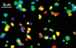
Nie's research team discovered a way to capture the quantum dots in specific quantities and in a wide range of colors and various intensities. The researchers made porous microbeads of polystyrene as small as 1 nm in diameter and seeded them with zinc sulfide-capped (ZnS) cadmium selenide (CdSe) nanocrystals (see Fig. 2). Both beads and quantum dots were made water-repellent to encourage the quantum dots to move into the pores. Once the quantum dots infiltrated the pores, they were sealed in. In ultraviolet light, each dot radiates a brilliant color. Novel optical properties, such as size-tunable emission and simultaneous excitation, also make the highly luminescent quantum dots ideal fluorophores for wavelength-and-intensity multiplexing.

To demonstrate the use of these quantum dots in DNA analysis, the researchers prepared microbeads of three colors, or spectral wavelengths, and attached them to strips of genetic material. Each color corresponded with a specific DNA sequence. These were used as probes to seek out complementary pieces of genetic material in a DNA mixture. The results confirmed that coding and target signals can be simultaneously read at the single-bead level.

Imaging and spectroscopic measurements indicate that the quantum dot-tagged beads are highly uniform and reproducible, yielding bead identification accuracies as high as 99.99% under favorable conditions. The researchers believe that this spectral coding technology will open new opportunities in gene expression studies, high-throughput screening, and medical diagnostics.

REFERENCE

  1. M. Han et al., Nature 19, 7 (2001).
About the Author

Sally Cole Johnson | Senior Technical Editor

Sally Cole Johnson has worked as a writer for over 20 years, covering physics, semiconductors, electronics, quantum, the Internet of Things (IoT), optics, photonics, high-performance computing, IT networking and security, neuroscience, and military embedded systems. She served as an associate editor for Laser Focus World in the early 2000s, and rejoined the editorial team as senior technical editor in January 2022.

Sponsored Recommendations

Request a quote: Micro 3D Printed Part or microArch micro-precision 3D printers

April 11, 2024
See the results for yourself! We'll print a benchmark part so that you can assess our quality. Just send us your file and we'll get to work.

Request a Micro 3D Printed Benchmark Part: Send us your file.

April 11, 2024
See the results for yourself! We'll print a benchmark part so that you can assess our quality. Just send us your file and we'll get to work.

Request a free Micro 3D Printed sample part

April 11, 2024
The best way to understand the part quality we can achieve is by seeing it first-hand. Request a free 3D printed high-precision sample part.

How to Tune Servo Systems: The Basics

April 10, 2024
Learn how to tune a servo system using frequency-based tools to meet system specifications by watching our webinar!

Voice your opinion!

To join the conversation, and become an exclusive member of Laser Focus World, create an account today!