Laser technology offers ultrafast x-ray imaging

Ron Olson
A new technique for producing medical x-rays promises increased imaging resolution while dramatically decreasing patient radiation exposure. The system is being pioneered by MXISystems (Nashville, TN) in collaboration with Positive Light (Los Gatos, CA). It uses a terawatt-class laser supplying 10-J, 8-ps pulses at 1 µm and an S-band linear accelerator (LINAC).
The laser beam collides with the electron beam in a target chamber producing 1- to 10-ps pulses via inverse Compton scattering at a nominal (adjustable) energy of 15 to 50 keV. The x-rays propagate in the direction of the electron beam and travel through a beryllium mirror as a collimated, spatially coherent pulse. Because the x-ray beam is diverging at <1°, the source can be directed to several different imaging locations, thus maximizing patient throughput and accelerating system payback.
The MXI system initially will carry a substantial price tag when compared to the cost of conventional x-ray machines (about ~$3 million). But according to company cofounder Marcus Mendenhall, a radiologist, the system price will fall to $1.5 million in production quantities and, moreover, will be justified independently by both improved image quality (implying improved diagnosis) and increased patient safety (from reduced x-ray exposure).
Imaging improvements
The improved resolution results from several source characteristics: the extremely short (2 to 10 ps) exposures eliminate motion as a concern (conventional x-ray exposures last for several hundred milliseconds); the ability to tune the x-rays allows optimal imaging for different tissue types (each with different absorption characteristics); and the monochromatic x-rays produce clearer images by avoiding broadband emissions (those outside the useful bandwidth) that add to the background noise without contributing to the signal. In addition, by using a new generation of gated detectors, MXI hopes to take advantage of time-of-flight (TOF) imaging not possible without an ultrafast x-ray source. The TOF x-rays will further enhance image clarity by eliminating noise produced by scatter within the tissues of the imaged body part.
Still another imaging improvement will involve phase-contrast radiography, enabled by the monochromatic x-rays. Phase-contrast imaging relies on differences in refractive indices of the various tissues rather than on the differences in absorption. Because the phase-shift coefficient is typically two to three orders of magnitude higher than the absorption coefficient at any given atomic number, it is theoretically possible to get a corresponding increase in information from the beam exiting the imaged tissue. Phase contrast works particularly well with low atomic number materials like carbon, hydrogen, and oxygen, which are the body's primary building blocks.
Laser light source
At the heart of the imaging system is a Ti:sapphire/Nd:glass laser system with chirped pulse amplification and an all-solid-state front end that was designed and built by Positive Light to assure extremely stable performance within a medical center/hospital environment. A Spectra-Physics (SP; Mountain View, CA) Millennia Nd:YVO3 laser pumps a SP Tsunami Ti:sapphire oscillator, tuned to 1054 nm, to produce a stream of 150-fs pulses. The pulses are temporally stretched to 1 ns in the Spitfire regenerative-amplifier stage pumped by a diode-pumped Evolution Nd:YLF laser supplying the regenerative amplifier with 10-mJ, 527-nm pump pulses at a rate of 1 kHz. Although the system repetition rate will be limited effectively to single-shot (~0.01 Hz) status by the final Nd:glass amplifiers, the diode-pumped 1-kHz regenerative stage offers the advantage of producing extremely stable seed pulses when compared to previous flashlamp-pumped designs.
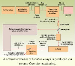
The 0.5-mj pulses are split via a beamsplitter (see figure on p. 165). Beam A is frequency-quadrupled to supply a 263-nm source to the fourth generation LINAC photocathode designed by Brookhaven National Laboratory (Upton, NY) to produce 1-nC electron bunches of 8-ps duration. The LINACs are being provided by Advanced Energy Systems (Medford, NY). Beam B is image relayed through a chain of Nd:phosphate-glass amplifiers resulting in single, 200-ps pulses with 20 J at 1054 nm. The pulses are then recompressed to 1 to 10 ps by a pair of gratings. The final 10-J output exhibits nearly diffraction-limited performance that is critical to the inverse Compton scattering process.
The new generation of laser systems is expected to mesh nicely with modern medicine. The technology has changed dramatically since Positive Light delivered its first terawatt laser in 1992, according to CEO Jeremy Weston. The company now implements all solid-state components to perform the critical beamshaping and preamplifier functions followed by a large aperture, relay-imaged power amplifier chain, he said.
Although the laser wavelength is fixed, the x-ray energy can be tuned between 15 and 50 keV by changing the RF drive power to the LINAC. An increased voltage corresponds to a more energetic collision between the electrons and the laser pulse within the target chamber, resulting in shorter-wavelength x-rays.
Targeting applications
While awaiting clinical trials and US Food and Drug Administration approval, MXISystems will market the system for related applications including small-animal imaging. Mendenhall has already been contacted by a NASA research group that is interested in studying effects of weightlessness, such as bone loss. The MXI system will allow repeated high-resolution x-rays to chart this condition on a relatively small number of living animals, thereby avoiding the hundreds and sometimes thousands of dissected mice that are typically sacrificed in such experiments.
Other targeted applications include industrial radiography to examine the dynamics within rocket engines. Because of the low divergent nature of the x-ray beam, the source can be tens of meters removed from the rocket enginesafely out of harm's way in the unlikely event of an explosion.
Industrial applications not withstanding, the system designers get most excited when the conversation turns back to medicine and specifically to the area of mammography. The ultrafast exposures are expected to provide more accurate images, alleviate patient discomfort caused by repeated imaging, and cut x-ray exposure by a factor ranging from 2 to 50 (depending on whether conventional or three-dimensional images are obtained). Consequently, the technology is expected to be welcomed by radiologists as well as the millions of women worldwide who receive mammograms on an annual basis.
RON OLSON, is a cofounder of Positive Light, Los Gatos, CA,; e-mail: [email protected].