Optical absorption imaging shows shadow of single atom in visible light
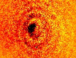
Brisbane, Australia--Scientists at Griffith University have captured images of the shadow cast by a single ytterbium atom in near-UV (369.5 nm) light. Because the characteristics of such a shadow can also easily be calculated, the technique is an effective way to test the limits of absorption imaging.
"We have reached the extreme limit of microscopy; you cannot see anything smaller than an atom using visible light," says Dave Kielpinski of Griffith University's Centre for Quantum Dynamics. "We wanted to investigate how few atoms are required to cast a shadow and we proved it takes just one." The results were published this week in Nature Communications.
The atom is ionized and held in a radio-frequency trap. The resulting image had a contrast of 3.1%—matching theoretical calculations. Kielpinski notes that if the frequency of the light shone on the atom is changed by just one part in a billion, the image can no longer be seen.
There are potential follow-on benefits for biomicroscopy. "Because we are able to predict how dark a single atom should be, as in how much light it should absorb in forming a shadow, we can measure if the microscope is achieving the maximum contrast allowed by physics," says Erik Streed, a member of the research team. "This is important if you want to look at very small and fragile biological samples such as DNA strands, where exposure to too much UV light or x-rays will harm the material. We can now predict how much light is needed to observe processes within cells, under optimum microscopy conditions, without crossing the threshold and destroying them."
Source: http://www3.griffith.edu.au/03/ertiki/tiki-read_article.php?articleId=37742
About the Author

John Wallace
Senior Technical Editor (1998-2022)
John Wallace was with Laser Focus World for nearly 25 years, retiring in late June 2022. He obtained a bachelor's degree in mechanical engineering and physics at Rutgers University and a master's in optical engineering at the University of Rochester. Before becoming an editor, John worked as an engineer at RCA, Exxon, Eastman Kodak, and GCA Corporation.