Compressed sensing allows super-resolution single-molecule microscopy imaging of live cell structures
Atlanta, GA--Researchers from the Georgia Institute of Technology and the University of California San Francisco (UCSF) have pushed the limits of super-resolution microscopy, using single-molecule identification at higher speeds to track live cells in action. By identifying many individual molecules at once using compressed sensing, this new method provides needed spatial resolution plus a faster temporal resolution than previously possible.
Now, Lei Zhu of Georgia Tech and and Bo Huang at UCSF have developed an advanced approach using super-resolution microscopy to resolve cellular features an order of magnitude smaller than what could be seen before. (The research was published April 22, 2012 in the journal Nature Methods.) Previous technology using a single-molecule-switching approach for super-resolution microscopy depends on spreading single molecule images sparsely into many, often thousands of, camera frames. It is extremely limited in its temporal resolution and does not provide the ability to follow dynamic processes in live cells.
"We can now use our discovery using super-resolution microscopy with seconds or even subsecond temporal resolution for a large field of view to follow many more dynamic cellular processes," says Zhu. "Much of our knowledge of the life of a cell comes from our ability to see the small structures within it." Huang noted, "One application, for example, is to investigate how mitochondria, the power house of the cell, interact with other organelles and the cytoskeleton to reshape the structure during the life cycle of the cell."
"The diffraction limit has long been regarded as one of the fundamental constraints for light microscopy until the recent inventions of super-resolution fluorescence microscopy techniques," says Zhu. Super-resolution microscopy methods, such as stochastic optical reconstruction microscopy (STORM) or photoactivated localization microscopy (PALM), rely on the ability to record light emission from a single molecule in the sample. Using probe molecules that can be switched between a visible and an invisible state, STORM/PALM determines the position of each molecule of interest. These positions ultimately define a structure.
The new finding is significant, said Zhu and Huang, because they have shown that the technology allows for following the dynamics of a microtubule cytoskeleton with a three-second time resolution, which would allow researchers to study the active transports of vesicles and other cargos inside the cell. Using the same optical system and detector as in conventional light microscopy, super-resolution microscopy naturally requires longer acquisition time to obtain more spatial information, leading to a trade-off between its spatial and temporal resolution. In super-resolution microscopy methods based on STORM/PALM, each camera image samples a very sparse subset of probe molecules in the sample. An alternative approach is to increase the density of activated fluorophores so that each camera frame samples more molecules. However, this high density of fluorescent spots causes them to overlap, invalidating the widely used single-molecule localization method.
The authors said that a number of methods have been reported recently that can efficiently retrieve single-molecule positions even when the single fluorophore signals overlap. These methods are based on fitting clusters of overlapped spots with a variable number of point-spread functions (PSFs) with either maximum likelihood estimation or Bayesian statistics. The Bayesian method has also been applied to the whole image set.
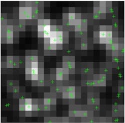
As a result of new research, Zhu and Huang present a new approach based on global optimization using compressed sensing, which does not involve estimating or assuming the number of molecules in the image. They show that compressed sensing can work with much higher molecule densities compared to other technologies and demonstrate live cell imaging of fluorescent protein-labeled microtubules with three-second temporal resolution. The STORM experiment used by the authors, with immunostained microtubules in Drosophila melanogaster S2 cells, demonstrated that nearby microtubules can be resolved by compressed sensing using as few as 100 camera frames, whereas they were not discernible by the single-molecule fitting method. They have also performed live STORM on S2 cells stably expressing tubulin fused to mEos2.
At the commonly used camera frame rate of 56.4 Hz, a super-resolution movie was constructed with a time resolution of three seconds (169 frames) and a Nyquist resolution of 60 nm, much faster than previously reported, said Zhu and Huang.
About the Author

John Wallace
Senior Technical Editor (1998-2022)
John Wallace was with Laser Focus World for nearly 25 years, retiring in late June 2022. He obtained a bachelor's degree in mechanical engineering and physics at Rutgers University and a master's in optical engineering at the University of Rochester. Before becoming an editor, John worked as an engineer at RCA, Exxon, Eastman Kodak, and GCA Corporation.