Only on the Web: MEDICALWATCH: Terahertz technology may improve medical imaging
Advances in ultrafast and diode lasers are once again playing a role in improving the way disease is detected and diagnosed. Scientists at Toshiba Research Europe Ltd. (TREL; Cambridge, England) have developed a new imaging technique that uses terahertz waves to produce a wealth of diagnostic information. Initial studies have shown that terahertz pulse imaging (TPI) holds enormous promise for certain types of medical imaging and analysis, including early detection of dental caries, identification and diagnosis of periodontal disease, burn-depth assessment, and detection and assessment of skin cancer, including melanoma.
Traditional diagnostic techniques such as x-ray radiography, computed tomography, magnetic-resonance imaging (MRI), and ultrasound penetrate deeply into the body and provide detailed renderings of various organs and tissues. However, terahertz waves offer imaging and diagnostic capabilities not afforded by conventional imaging modalities. In addition, they are considered safer than x-rays because the waves are nonionizing, and low average powers are used; the technology has the potential to be less expensive than more-complex imaging modalities such as MRI. "These are early days, but it is clear that TPI is going to be very important, particularly in areas where x-rays are insensitive," says Michael Pepper, managing director of the TREL Cambridge Research Laboratory.
The TPI system developed at TREL represents a safe, noninvasive, inexpensive clinical imaging modality with additional diagnostic capabilities, according to Dr. Don Arnone, TPI project leader. Its particular advantages include the ability to distinguish between healthy and abnormal soft tissue, to analyze the internal composition of tissue samples, and to build a three-dimensional (3-D) digital image using the acquired data.
TPI also appears to solve at least one significant problem with laser-based imaging techniques such as fluorescence imaging and optical coherence tomography, which operate in the near-infrared and visible regions. The development of optical-imaging methods has been hampered by the scattering of photons in tissue at wavelengths in the near-IR and visible regions (400-1300 nm). For highly scattering or thick media, image-bearing light such as ballistic or snake photons become weak, making image reconstruction much more complicated and data intensive. In contrast, terahertz energy has very long wavelengths (15 µm-3 mm), which can significantly reduce light scattering and improve image quality and signal-to-noise ratios. "The ultimate goal of TPI is to provide high-quality images that contain diagnostic information not readily available with other techniques," Arnone says.
Ti:sapphire source
Medical researchers have long been interested in tapping into this potential but were stymied by technological and cost barriers when trying to generate and detect radiation in the terahertz range (between infrared and micro wave). Fortunately, recent developments in pulsed-laser and near-infrared diode-laser technology have opened the door to the exploitation of this frequency range. Terahertz waves can now be generated efficiently by illuminating a semiconductor crystal with ultrafast pulses of laser energy.
FIGURE 1. The entire TPI system is controlled by a desktop personal computer. To image an object, it is mounted on the rear of the terahertz-generation crystal; both the object and the generation crystal are then stepped through the visible generation beam. An x-y translation stage allows terahertz spectra to be recorded at each pixel in the image. Larger objects and those not easily accessible or easily mounted on the generation crystal can be coupled to the system using more sophisticated terahertz and/or visible optics, making it possible to perform TPI on both large and small objects in vivo.
The TREL system comprises a femto second pulsed visible laser, a terahertz generator, coupling optics for the terahertz beam, and time-resolved electro-optical sampling (EOS) measurement of the terahertz pulses (see Fig. 1). The visible laser source is a diode-pumped Ti:sapphire laser oscillator tunable in the 750-890-nm range, producing short pulses down to 50 fs with a maximum power of 1 W. The terahertz waves are generated using difference-frequency mixing in a semiconductor crystal (zinc telluride; ZnTe) by focusing the visible beam to a 250-µm-diameter spot using a concave mirror. A beamsplitter enables the visible laser beam to both generate and detect the terahertz waves using an EOS detection crystal (also ZnTe).
The system's ability to operate as both an imaging device and a spectroscopic-analysis tool makes it unique among currently available medical-imaging techniques. For example, used in time-of-flight mode to visualize internal structures, the system can build a 3-D image from transmitted signals. The target is illuminated by a pulsed terahertz wave, most of which passes right through, but some of which is reflected from the interfaces between internal layers. The delay of the transmitted pulse as it passes through the object gives a precise measure of the distance to the various surfaces inside the object. By scanning the beam across the target, a complete 3-D picture of the internal structure can be created, and the resulting image can be displayed and manipulated as desired on-screen.
When operated as a spectroscope, the system can determine an object's composition at any point. In this mode, the object under examination is illuminated by a broad frequency band of terahertz waves. A detector examines how the various frequencies are absorbed or dispersed by the object. The resulting absorption spectrum provides a characteristic signature or fingerprint for that material. Time-domain data are displayed on the computer screen, or frequency-domain data can be examined at each pixel in the image by applying a fast Fourier transform to the time-domain data.
This ability to provide the terahertz spectral fingerprint of the object at each pixel in its image is one of the major advantages of TPI relative to x-ray, MRI, and other conventional imaging techniques, according to Arnone. Spatial resolution of 200 µm is common using the current version of the TREL TPI system, and early studies have shown that different soft-tissue types may have characteristic spectral fingerprints in the terahertz range.
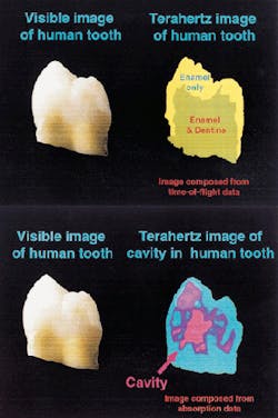
FIGURE 2. Two-dimensional contour plot of the time-of-flight data at each value of x and y shows how TPI can be used to map regions of enamel and enamel plus dentine and accurately determine the thickness of each region (top). The same terahertz data set can be manipulated to produce an image of the pulp cavity. Note the strong absorption in the pulp cavity, which appears to reflect the buildup of additional material (bottom).
Initial applications
Dental imaging is one of the first TPI applications being studied by the TREL team. Early results suggest that this technique may provide valuable diagnostic information about the enamel, dentine, and pulp cavity of a tooth (see Fig. 2). In one mode, the researchers were able to measure the thickness of tooth enamel on the outside, while in another mode they displayed the internal tissue or dentine, highlighting the condition of the tooth cavity. In addition, they were able to construct a 3-D image of the tooth and rotate it on the computer screen-a feature dentists may one day use to examine each tooth from the optimum angle.
"Thus far this has only been done for an extracted tooth, but we hope to develop the system so that it can be used on patients in the dentist's chair in the near future," Arnone says. Initial studies are expected to focus on the ability to characterize teeth with primary and secondary caries and to detect periodontal disease in live teeth. Preliminary development of an in vivo instrument to image teeth using terahertz waves is also under way.
The TREL team is also working to overcome the issue of image-acquisition time, which is now on the order of hours for teeth. According to Arnone, this is due primarily to the fact that initial studies were done using a low-power (1 mW) system, and TREL researchers have already begun developing more-efficient radiation sources.
"Combining these with more-powerful pump lasers means that much higher powers are now available," Arnone says. "We hope to get the acquisition time down to minutes (or less) for teeth over the next several months." Researchers are also working to improve penetration depth, which would help make the technology more competitive with conventional imaging methods, and to develop a more compact system that would be more clinically practical and cost-effective.
At this point, Arnone anticipates beginning preclinical trials for dental imaging late next year and clinical trials another 18 months to two years after that. In addition, preliminary measurements on TPI of different animal soft-tissue types have also been taken, although further study is required to confirm the usefulness of terahertz imaging in the diagnosis of diseased or affected tissue in these areas, according to Arnone. In the meantime, TREL is interested in discussing possible commercialization of this technology with interested parties.
